

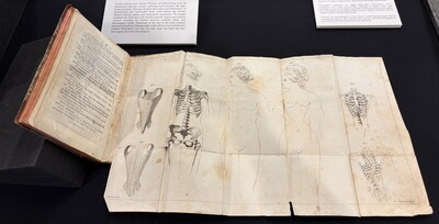
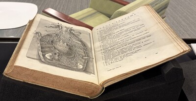
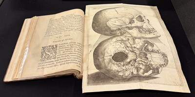
Between the sixteenth and eighteenth centuries, the study of human anatomy was transformed by the printed image. Long before modern medical technologies allowed physicians to look inside the body, anatomical knowledge depended on careful observation through dissection and on the ability to record those observations visually. With the rise of printing, woodcut and engraved illustrations became essential tools for studying and communicating the structure of the human body.
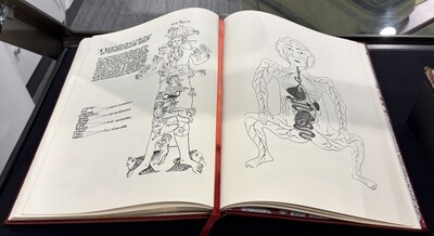
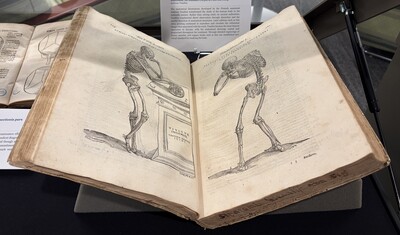
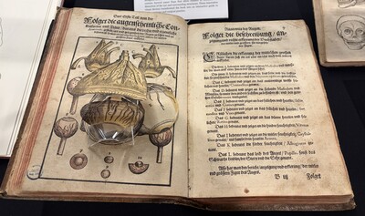
The works in this collection trace the development of anatomical illustration during this formative period. Early printed anatomical texts, such as the work of Johann Dryander, reflect a moment of transition when traditional medical knowledge began to intersect with new methods of observation. Only a few years later, the publications of Andreas Vesalius revolutionized the study of anatomy by grounding it in what could be directly seen during dissection. His richly detailed illustrations established a new visual language for representing the body.
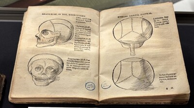
Later authors expanded and refined this approach. Anatomists such as Juan Valverde de Amusco and Pieter Paaw adapted Vesalian methods to focus on specific structures of the body, while eighteenth-century physicians like Samuel Thomas von Sömmerring applied anatomical illustration to new medical questions. Together, these works show how artists, printers, and physicians collaborated to make the invisible structures of the human body visible.
For additional questions about this collection, contact an archivist at 713-799-7145 or mcgovern@library.tmc.edu
 by Samuel Thomas von Sömmerring")


